Dr. Sohrab Ali
Endourology & Kidney Disease Specialist
BOOK APPOINTMENT

Meet
Dr. Sohrab Ali
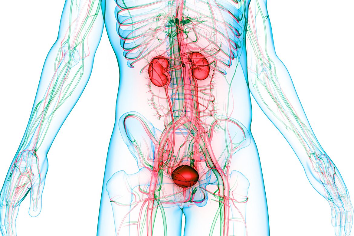
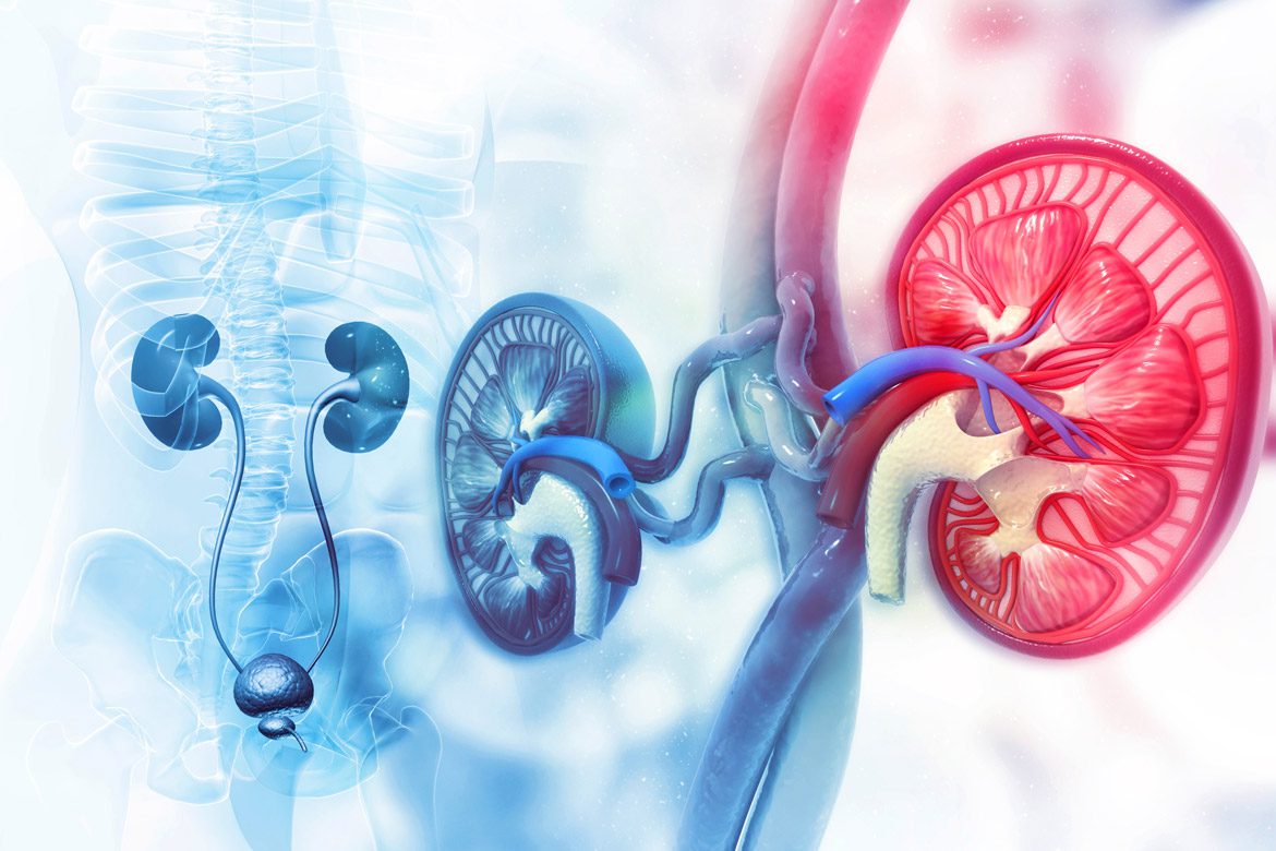
Dr. Sohrab Ali is a robotic and minimally invasive surgeon in Orange County, CA. He serves as an Assistant Clinical Professor at the University of California, Irvine specializing in Endourology and Minimally Invasive Oncology. An expert in robotic partial and radical nephrectomy, robotic prostatectomy, and percutaneous nephrolithotomy (PCNL), he specializes in the retroperitoneal approach and single-port robotics.
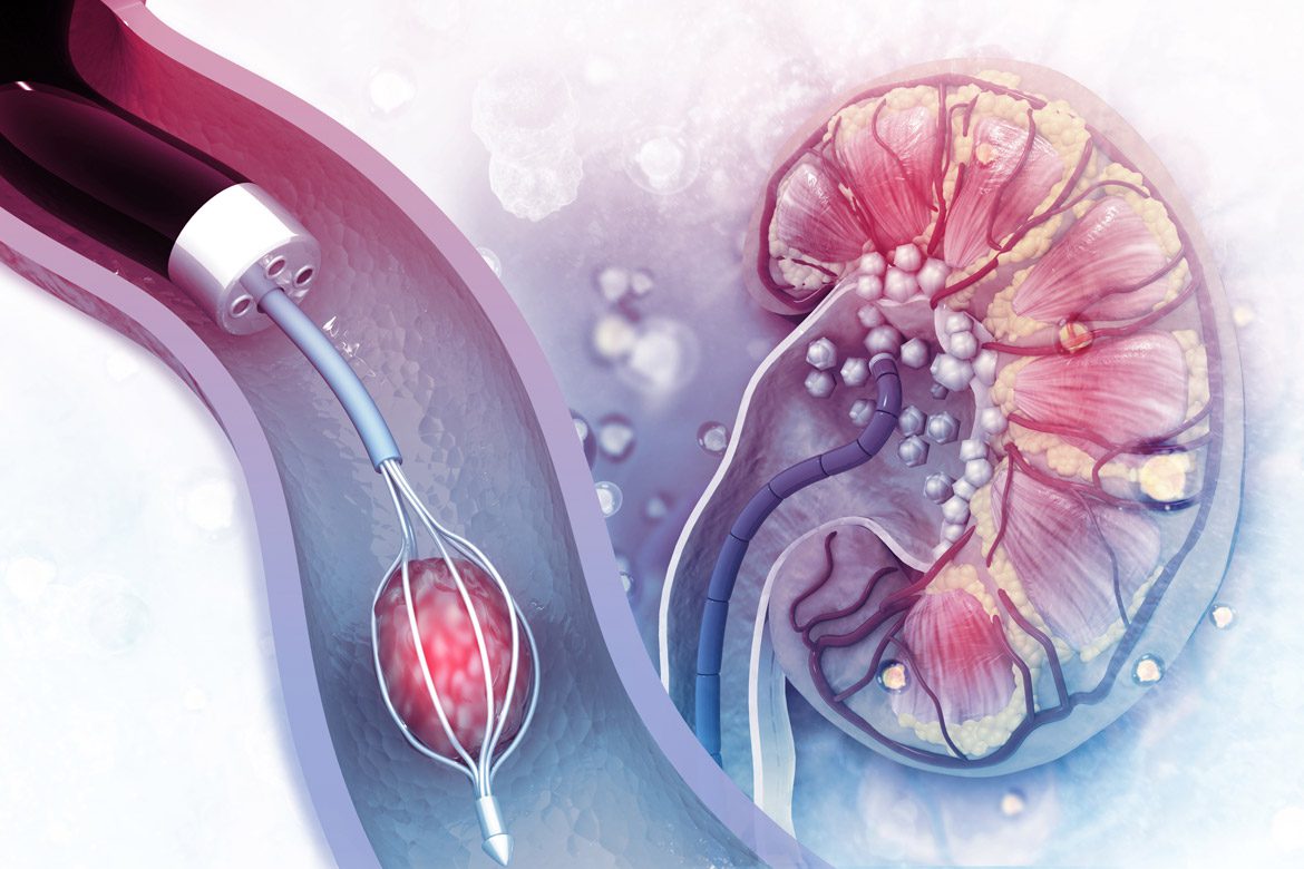
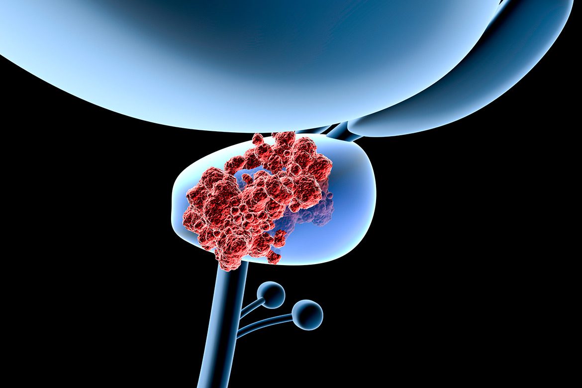
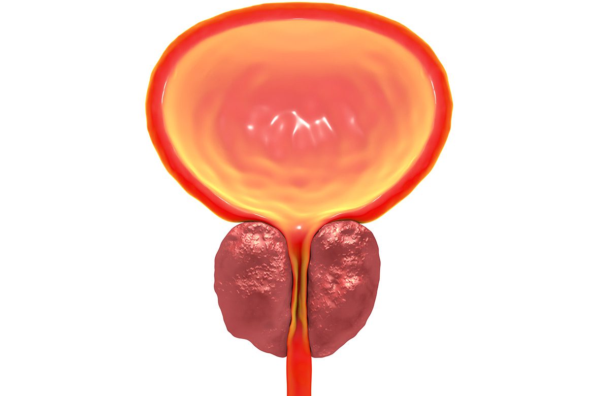
As a kidney stone and disease specialist, he routinely manages complex stone disease. He is an active researcher in the Curiosity and Innovation laboratory, including PI and Co-PI on multiple clinical trials including the use of baking soda for stone prevention and dissolution in calcium oxalate and uricacid stone formers, plus the use of Indocyanine-green for mapping and preserving neuro-vascular bundles during radical prostatectomy. Dr. Ali is also a BPH specialist. He also serves as the director of the UCI Urology Grandest Rounds program.
Specialties


Testimonials
Having Dr. Ali for my kidney stone removal process was a very pleasant experience, both in receiving clear and concise easy to understand explanations of conditions and causes, and in preparation for procedure and aftercare preventative maintenance.
Everyone who works there is very nice and helpful. Dr. Ali is the best!! I feel so much better now. Thank you so much for saving my kidney!!
Dr. Ali was excellent in dealing with my prostate cancer. He did a robotic procedure. He guided me thru decisions prior to my decision to go for surgery. He did an excellent surgery, as my recovery was near painless and I was feeling very good within days.