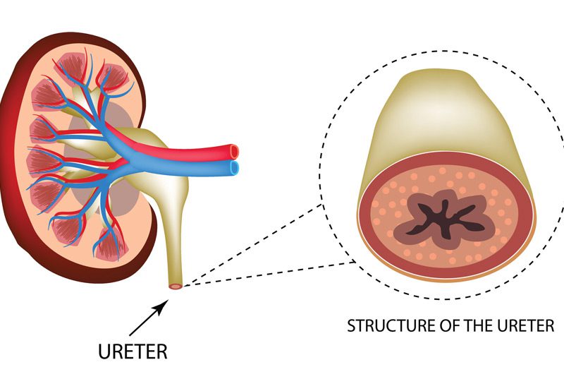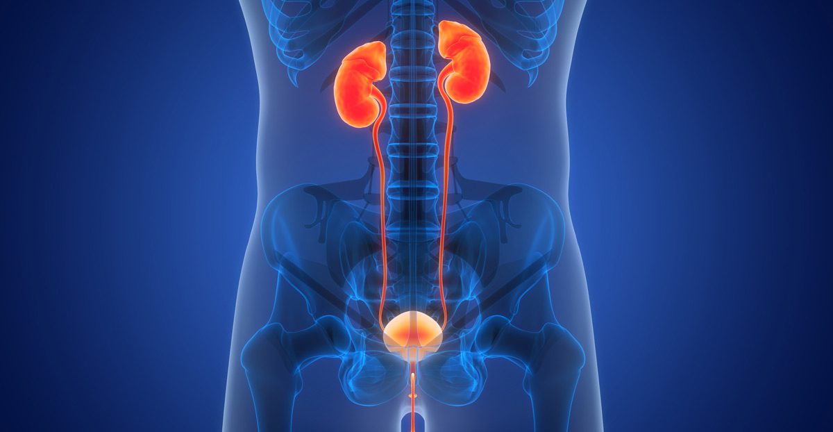

Ureteropelvic junction (UPJ) obstruction is a condition in which the flow of urine is impaired at the junction where the kidney meets the ureter—the narrow tube that carries urine to the bladder. This blockage can lead to urine buildup within the kidney, causing swelling (hydronephrosis), discomfort, and over time, reduced kidney function. UPJ obstruction can be congenital or acquired and may affect one or both kidneys.
Symptoms of UPJ Obstruction
Not all patients with UPJ obstruction experience noticeable symptoms, especially in the early stages. When symptoms do occur, they may include:
- Flank or abdominal pain, especially after fluid intake
- Nausea or vomiting
- Recurrent urinary tract infections (UTIs)
- Presence of blood in the urine (hematuria)
- Kidney stones
- Decreased kidney function over time

Causes and Risk Factors
UPJ obstruction can be present at birth or develop later due to structural changes, scarring, or external pressure on the ureter. Common causes include:
- Congenital Narrowing: Incomplete development of the junction before birth
- Crossing Blood Vessels: Blood vessels pressing against the ureter and causing compression
- Scar Tissue: Often resulting from previous surgery, infection, or trauma
- Ureteral Kinks or Adhesions: That interfere with urine flow
- Tumors or External Masses: Rarely, tumors can compress the UPJ from outside
Diagnosis
Timely diagnosis is critical to prevent long-term kidney damage. Evaluation typically includes:
- Ultrasound: To detect hydronephrosis or kidney enlargement
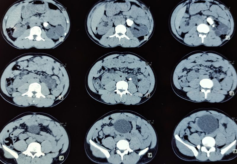
- CT Urogram or MRI: To identify the exact location and cause of the obstruction
- Nuclear Renal Scan (Lasix Renogram): Measures how well each kidney functions and how effectively it drains urine
- Voiding Cystourethrogram (VCUG): Sometimes used to rule out reflux or other anatomical abnormalities
Treatment Options
The goal of treatment is to relieve the obstruction, preserve kidney function, and alleviate symptoms.
- Observation: In mild or asymptomatic cases, regular monitoring may be appropriate
- Pyeloplasty: The most common surgical treatment. This involves removing the narrowed segment and reconnecting the healthy portions of the kidney and ureter. It can be performed using:
- Robotic-Assisted Pyeloplasty: Minimally invasive, with less pain, smaller incisions, and faster recovery
- Laparoscopic or Open Pyeloplasty: Depending on the complexity of the obstruction
- Endopyelotomy: A less-invasive internal incision of the stricture using a scope, typically reserved for select patients
- Temporary Drainage: In some cases, a nephrostomy tube or stent may be used to relieve pressure prior to surgery
Next Steps
If you’re experiencing persistent flank pain, recurrent infections, or have been diagnosed with hydronephrosis or kidney dysfunction, a thorough evaluation by a urologist is essential. UPJ obstruction can often be corrected with a minimally invasive procedure that restores normal drainage and protects long-term kidney health.
