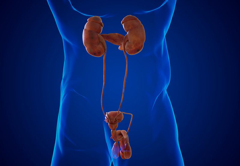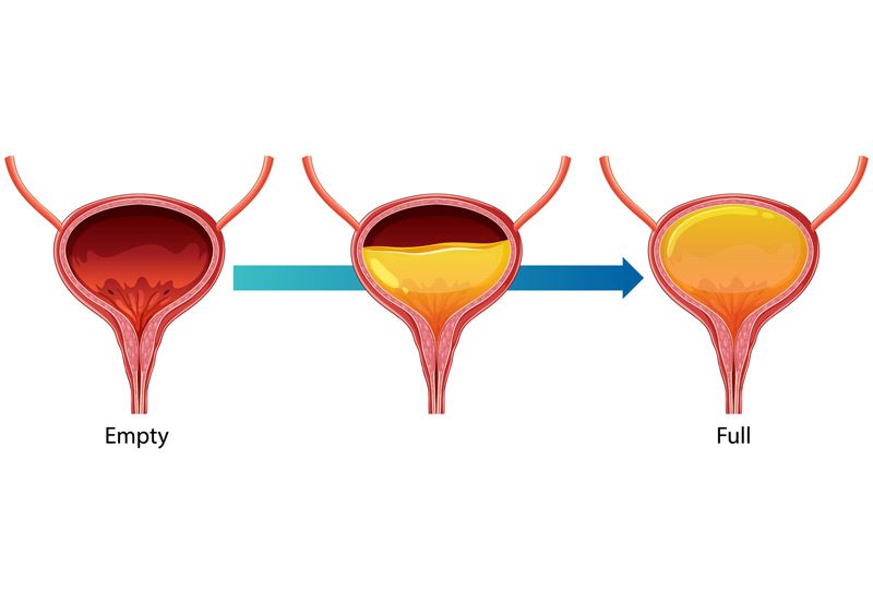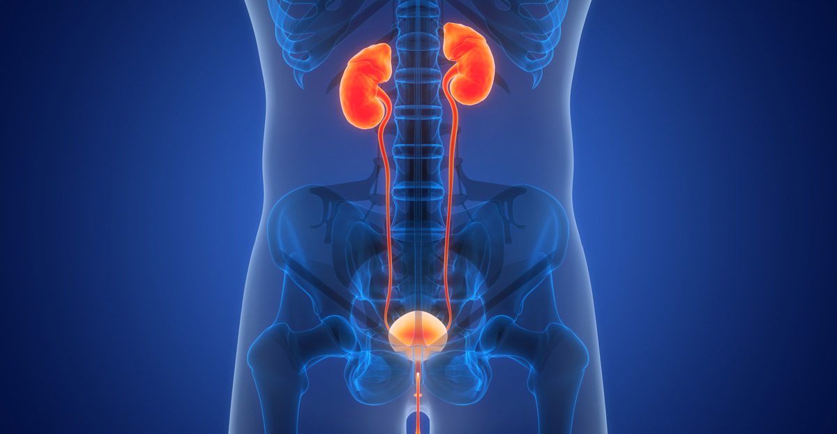

Ureteral reimplantation is a surgical procedure designed to correct abnormalities in the connection between the ureter and the bladder. It is most commonly performed to treat vesicoureteral reflux (VUR), a condition where urine flows backward from the bladder into the ureters or kidneys, or to address ureteral obstruction or injury that impairs the normal flow of urine. By repositioning the ureter into the bladder wall, this procedure restores proper urinary drainage and helps prevent future infections and kidney damage.
Causes of Ureteral Abnormalities
Several conditions may lead to the need for ureteral reimplantation. Vesicoureteral reflux is often congenital, caused by a defect in the natural valve that prevents backflow of urine. Ureteral obstruction can occur due to scarring from previous surgeries, kidney stones, chronic infections, or injury to the ureter. In some cases, tumors or anatomical abnormalities may compromise the ureter’s function and require surgical correction.

Diagnosis
A thorough diagnostic process is necessary before considering ureteral reimplantation. Common evaluations include:
- Imaging Tests: CT urograms, renal ultrasounds, or MRI may be used to assess the anatomy of the urinary tract
- Voiding Cystourethrogram (VCUG): Often used to confirm vesicoureteral reflux
- Nuclear Medicine Renal Scan: Evaluates kidney function and drainage
- Cystoscopy: Direct visualization of the bladder and ureteral openings
These tests help determine the location, severity, and cause of the ureteral problem and guide surgical planning.
Treatment Options
- Open Ureteral Reimplantation: The surgeon makes a small incision in the lower abdomen to access the bladder. The affected ureter is detached from its abnormal location and reimplanted into a new position within the bladder wall to create a longer submucosal tunnel. This helps restore the one-way valve mechanism.
- Minimally Invasive Approaches: In selected cases, laparoscopic or robotic-assisted techniques may be used to perform the procedure with smaller incisions and a quicker recovery.
- Stenting: A temporary ureteral stent may be placed during surgery to aid healing and ensure urine flows properly during recovery.
Next Steps
Recovery typically involves a short hospital stay, catheter placement, and pain management. Most patients can return to normal activities within a few weeks. Follow-up imaging—such as a renal ultrasound or VCUG—is often recommended to confirm proper ureteral positioning and resolution of reflux or obstruction. Ongoing monitoring ensures long-term protection of kidney function and urinary health.
