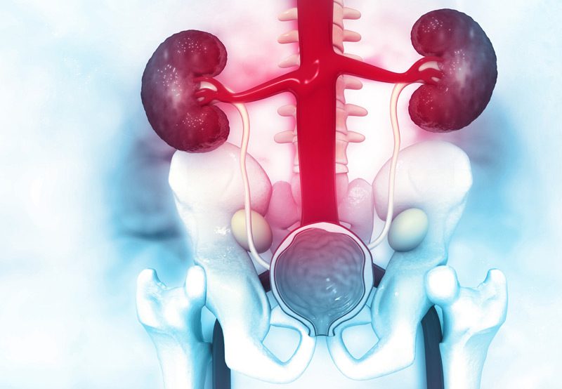

Laser endopyelotomy is a minimally invasive surgical procedure used to treat ureteropelvic junction (UPJ) obstruction—a condition in which urine flow is partially or completely blocked at the junction where the ureter meets the kidney. This obstruction can lead to symptoms such as flank pain, urinary tract infections, and progressive damage to kidney function. Laser endopyelotomy is an alternative to more invasive surgical reconstructions and is typically considered in select patients based on anatomical and functional considerations.
Symptoms of UPJ Obstruction
- Flank or back pain, often intermittent
- Recurrent urinary tract infections
- Nausea and vomiting associated with fluid buildup
- Visible or microscopic blood in the urine (hematuria)
- Reduced kidney function or hydronephrosis on imaging

Causes of UPJ Obstruction
UPJ obstruction may be:
- Congenital: Present from birth due to abnormal development or due to a crossing vessel which may compress and obstruct the ureter.
- Acquired: Resulting from scar tissue, previous surgery, kidney stones, or inflammation
Diagnosis
A thorough evaluation is essential before considering laser endopyelotomy. Diagnostic tools may include:
- Renal ultrasound or CT scan: To assess hydronephrosis
- Nuclear renal scan: To evaluate kidney function and drainage
- Retrograde pyelogram or CT urogram: To visualize the ureter and obstruction
- Cystoscopy with ureteroscopy: May be used for direct visualization during planning
Laser Endopyelotomy Procedure
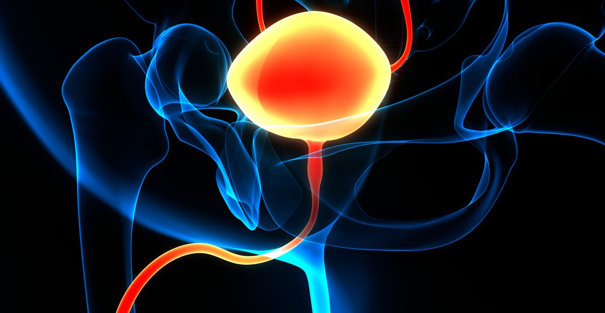
The procedure is performed using a ureteroscope, which is inserted through the urethra and bladder to access the ureter and kidney.
Once the area of narrowing at the UPJ is identified, a laser—typically Holmium:YAG or Thulium—is used to make a precise incision through the scarred or obstructed tissue.
- Balloon dilation may be used in conjunction with the laser to expand the narrowed segment.
- Stent placement: A ureteral stent is placed to maintain patency during the healing process and is usually left in place for 4–6 weeks.
The entire procedure is done under general anesthesia and usually takes about 1–2 hours.
Recovery and Success Rates
Patients can typically go home the same day or after an overnight observation. Most return to normal activity within a few days.
Follow-up imaging is performed after stent removal to ensure the obstruction has resolved.
Laser endopyelotomy offers good outcomes in selected patients, especially those with short, simple strictures and preserved kidney function.
However, recurrence of obstruction is possible, and open or robotic pyeloplasty may be needed in such cases.
Next Steps
If you have been diagnosed with a ureteropelvic junction obstruction and are looking for a minimally invasive option,
laser endopyelotomy may be a suitable treatment.
A detailed consultation and evaluation with a urologist can help determine whether you are a good candidate
based on your anatomy, stone history, and kidney function.
