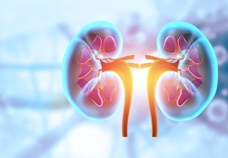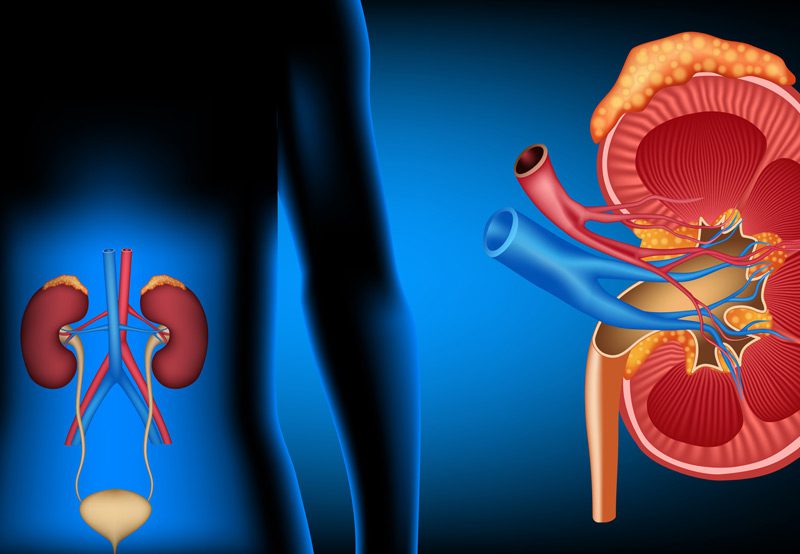

Pyeloplasty is a surgical procedure performed to correct ureteropelvic junction (UPJ) obstruction—a blockage at the junction where the kidney meets the ureter, the tube that carries urine to the bladder. When this connection is narrowed or blocked, it can impede urine flow, leading to pain, infection, kidney stones, or progressive kidney damage. Pyeloplasty restores proper drainage by removing the obstruction and reconstructing the junction.
Causes of UPJ Obstruction
UPJ obstruction can occur due to several factors, including:
- Congenital Narrowing: A birth defect causing a tight or underdeveloped junction
- Crossing Blood Vessels: Vessels compressing the ureter externally
- Scar Tissue or Previous Surgery: Adhesions or inflammation from past procedures
- Stones or Tumors: Rarely, obstructive lesions may be responsible
This condition may present with symptoms like flank pain, recurrent urinary tract infections, kidney stones, or a sensation of fullness in the abdomen. In some cases, it may be detected incidentally through imaging for unrelated issues.

Diagnosis
A diagnosis of UPJ obstruction is typically made through:
- Ultrasound: Detects hydronephrosis (swelling of the kidney due to fluid buildup)
- CT Urography: Provides detailed anatomical imaging of the kidneys and urinary tract
- MAG3 Renal Scan: Measures kidney function and drainage, confirming the severity of obstruction
- MRI or IV Pyelogram: May also be used to further define anatomy
These studies help determine if surgery is necessary based on the degree of obstruction and kidney function.
Treatment Options
The gold standard for treating UPJ obstruction is pyeloplasty. When performed robotically or laparoscopically, the procedure includes:
- Minimally Invasive Access: Small incisions for robotic instruments and camera
- Excision of Blocked Segment: The narrowed part of the ureter is removed
- Reconstruction: The healthy ends of the renal pelvis and ureter are stitched together to create a wider, unobstructed passage
- Stent Placement: A temporary ureteral stent is often inserted to aid healing and ensure urine flow
Robotic pyeloplasty provides precise suturing and enhanced visualization, resulting in high success rates with minimal discomfort and faster recovery.
Next Steps
After surgery, patients typically stay in the hospital for 1–2 days. The ureteral stent remains in place for a few weeks and is removed during a follow-up visit. Most individuals can resume light activity within a few days and return to full activity in a few weeks. Follow-up imaging is essential to confirm restored drainage and preserve kidney function. With proper diagnosis and timely surgical correction, pyeloplasty offers excellent long-term outcomes.
