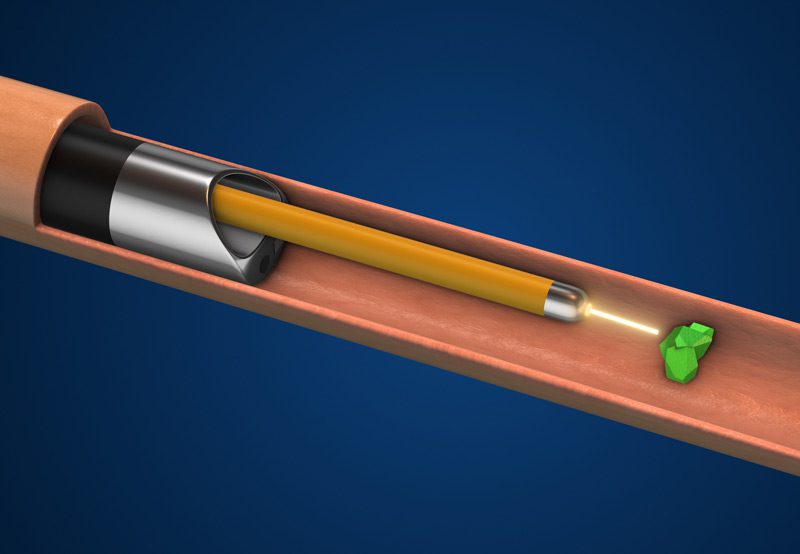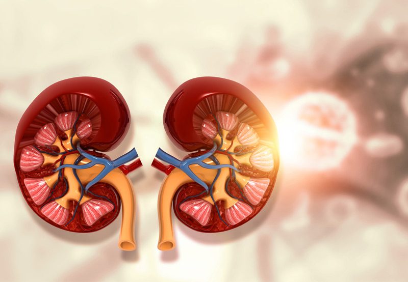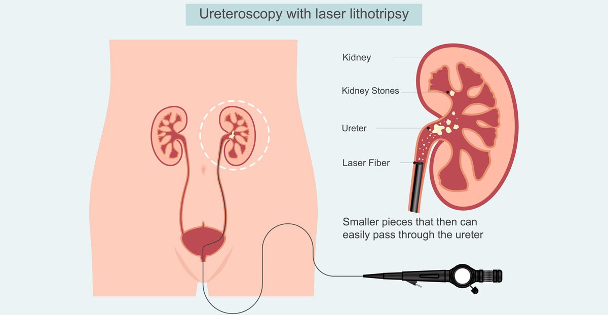

Ureteroscopy with lithotripsy is a minimally invasive procedure used to diagnose and treat stones located in the ureter or kidney. This approach combines two key components: a ureteroscope, a thin, flexible or rigid tube with a camera and light, and a lithotripsy device, which uses energy to break stones into small fragments that can be removed or passed naturally. It is often recommended when stones are too large to pass on their own, are causing significant pain or obstruction, or have not responded to other treatments like medication or shockwave therapy.
Causes of Stone Formation
Kidney and ureteral stones form when minerals and salts in the urine crystallize and stick together. Risk factors include dehydration, high dietary salt or protein intake, certain metabolic conditions, urinary tract infections, and family history of stones. If a stone travels into the ureter and becomes lodged, it can cause severe pain, infection, or obstruction of urine flow, requiring prompt intervention.

Diagnosis
Before performing ureteroscopy with lithotripsy, imaging tests are used to confirm the location, size, and type of stone:
- Non-contrast CT Scan: The gold standard for detecting stones
- Ultrasound: Useful for detecting larger stones and assessing kidney function
- X-rays or KUB (Kidney, Ureter, Bladder) Films: Sometimes used to monitor radiopaque stones over time
These tests help the urologist determine the best treatment approach and rule out other causes of symptoms.
Treatment Options
- Ureteroscopy with Laser Lithotripsy: The most common form uses a holmium:YAG laser to fragment stones into small pieces. The ureteroscope is passed through the urethra and bladder into the ureter without any incisions.
- Stone Removal: In some cases, stone fragments are directly extracted using a small basket tool inserted through the ureteroscope.
- Stent Placement: A temporary ureteral stent may be placed to promote healing, relieve swelling, and ensure continued urine flow after the procedure.
This approach is especially useful for mid- to lower-ureteral stones and for treating stones resistant to extracorporeal shock wave lithotripsy (ESWL).
Next Steps
Patients typically go home the same day and can resume light activities within 24–48 hours. Some discomfort during urination, minor bleeding, or increased urgency may occur temporarily. A follow-up visit is usually scheduled within 1–2 weeks to monitor healing, assess stone clearance, and remove the stent if placed. Preventive strategies, including dietary changes and metabolic evaluations, may be recommended to reduce the risk of future stone formation.
